M. Wang, J.R. Unruh, C. K. Johnson, R.L. Schowen, R.T. Borchardt and K. Kuczera.
Biochemistry 45:7778-7786 (2006).
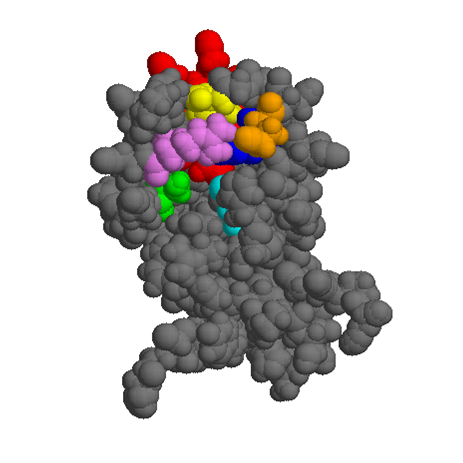
Domain motions of S-adenosyl-L-homocysteine hydrolase (AdoHcy hydrolase) have been detected by time-resolved fluorescence anisotropy measurements. Time constants for reorientational motions in the native enzyme were compared with those for enzymes where key residues were altered by site-directed mutation. Mutations M351P, H353A, and P354A were selected in a hinge region for motion between the open and closed forms of the enzyme, as identified in a previous normal mode study (Wang et al, Biochemistry 44:7228-7239, 2005). In wild type, substrate-free AdoHcy hydrolase (NAD+ cofactor in each subunit), reorientational motions were detected on time scales of 10-20 ns and 80-90 ns. The faster motion is attributed to domain motion and the slower motion to tumbling of the enzyme. Domain motion was also detected for the enzyme complexes E(NADH:3’-keto-adenosine) and E(NAD+:3’-deoxyadenosine), but was absent for the complex E(NADH:3’-keto-neplanocin A). The results indicate that AdoHcy hydrolase exists in equilibrium of open and closed structures, with the equilibrium shifted toward the more mobile open form for the substrate-free enzyme, E(NAD+), and for intermediates formed early in the catalytic cycle after substrate binding or late prior to substrate release, E(NAD+:ligand). However, the strong inhibitor neplanocin A upon binding undergoes oxidation forming the complex E(NADH:3’-keto-neplanocin). For this complex, which is analogous to the enzyme complex with the central catalytic intermediate, the equilibrium was shifted towards the more rigid closed form. A similar pattern was observed for M351P and P354A mutants. In contrast, domain motion could not be detected, either in the absence or presence of ligands or with the cofactor in either oxidized or reduced state, for the H353A protein, suggesting that this mutation changes the hinge-bending dynamics of the enzyme.
|
|
| SAHH catalytic cycle. | SAHH monomer - closed form with bound ligand. |
_______________________________________________________________________________
Q. Cheng, D. R. Benson, M. Rivera-Laos and K. Kuczera,
Biopolymers 83:297-312 (2006).
Two membrane-bound isoforms of cytochrome b5 have been identified in mammals, one associated with the outer mitochondrial membrane (OM b5) and the other with the endoplasmic reticulum (microsomal, or Mc b5). The soluble heme binding domains of OM and Mc b5 have highly similar three-dimensional structures but differ significantly in physical properties, with OM b5 exhibiting higher stability due to stronger heme association. In this study, we present results of 8.5 ns length molecular dynamics simulations for rat Mc b5, bovine Mc b5 and rat OM b5, as well as for two rat OM b5 mutants that were anticipated to exhibit properties intermediate between those of rat OM b5 and the two Mc proteins: the A18S/I32L/L47R triple mutant (OM3M) and the A18S/I25L/I32L/L47R/L71S quintuple mutant (OM5M). Analysis of the structure, fluctuations and interactions showed that the five b5 variants used in this study differed in organization of their molecular surfaces and heme binding cores in a way that could be used to explain certain experimentally observed physical differences. Overall, our simulations provided qualitative microscopic explanations of many of the differences in physical properties between OM and Mc b5 and two mutants in terms of localized changes in structure and flexibility. They also reveal that opening of a surface cleft between hydrophobic cores 1 and 2 in bovine Mc b5, observed in two previously reported simulations [Storch E.M. and Daggett V. Biochemistry (1995) 34:9682-9693; Altuve A, et al., Biochemistry (2001) 40:9469-9483], probably resulted from removal of crystal contacts and likely does not occur on the ns time scale. Finally, the MD simulations of OM5M b5 verify that stability and dynamic properties of cytochrome b5 are remarkably resistant to mutations that dramatically alter the stability and structure of the apoprotein.

|

|
| Surface cleft size in MD of cb5 isoforms. | Residues 25, 58, 71 and heme in bovine Mc cb5. | Residues 25, 58, 71 and heme in OM5M mutant. |